In 2014, I was in Wisconsin being trained in the Schroth Method for scoliosis. As we were going through the course, my instructor demonstrated a different way of imaging scoliosis. It took measurements automatically of posture and gave a 3D picture of the spine.
[video_player type=”youtube” style=”1″ dimensions=”560×315″ width=”560″ height=”315″ align=”center” margin_top=”0″ margin_bottom=”20″ ipad_color=”black”]aHR0cHM6Ly95b3V0dS5iZS9aanVVYlY3X1o2Yw==[/video_player]
At that time, I hadn’t started Align Therapy yet, but I knew I wanted to treat scoliosis with the best techniques and equipment available. That meant, I needed to figure out a way to get a Diers Formetric Surface Topography machine in house!
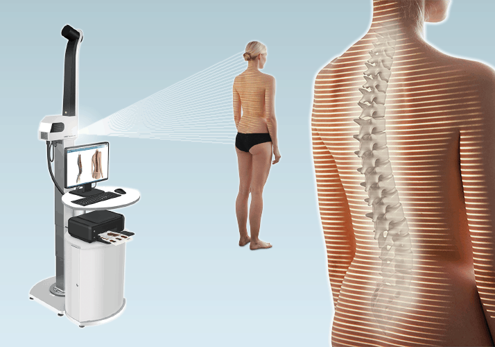
A year later, and after selling a kidney on the black market, my goal was realized when a huge box was delivered from Germany containing my very own Diers Formetric!
Since then, we have used this on hundreds of scoliosis and posture patients, and it has been a great tool to visualize, educate, and monitor these conditions. I can’t imagine treating scoliosis and spinal problems without this amazing tool.
Surface Topography has many advantages over traditional x-ray, but it also has some limitations.
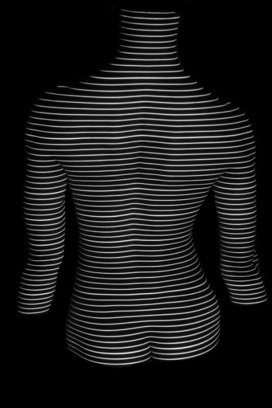
First, how does it work? Surface Topography is performed by shining lines of light on the back and then taking a picture. This picture is then analyzed with complicated math algorhythms that I don’t understand and a 3D representation of the spine is created.
In the past, measurements between the lines and calculations were made by hand. This was very time consuming and also had a higher error rate. With the Diers Formetric, this all happens automatically. After the picture is taken, the computer does all the calculations and measurements, usually without help.
We could go into more detail on the “magic” the machine performs, but you can go to Diers website to learn more.
Next, lets go over some of the advantages and disadvantages of Surface Topography.
Advantages
No X-Ray Radiation
All x-ray machines use radiation to create an image of the spine. These x-ray particles travel through the body and are absorbed more by bone than by soft tissue. This leaves an image behind of where the bones and other structures are. (This is a very simplistic view of x-ray)
With these images, the subject is exposed to radiation. Now, not all x-ray machines are created equal. There are many out there in smaller clinics and hospitals that have a large dose of raditation, but there are also low dose as well as ultra low dose x-ray machines out there as well that have a much lower exposure.
Kids with scoliosis are x-rayed on average 12+ times during their adolescent years. This increased exposure has been shown in the research to increase their cancer risk by 4 times.
Now, I am not saying they shouldn’t be imaged using x-ray, but it brings up the argument of whether there is a better way to monitor these curves. I am not anti-x-ray! In fact, I don’t usually treat someone unless I have their x-ray because I want to make sure there is not something odd going on in the spine.
My hope is that in the future we will be able to utilize surface topography to check for progression between follow ups with spine specialists. If we are not seeing changes on topography, then maybe we wait a bit before another x-ray, and limit exposure.
Automatic Measurements
One of the best things about the Diers Formetric is it automatically finds landmarks on the back. After it finds those landmarks it will then automatically make calculations and measurements and provide them in an easy to read and understand graph.
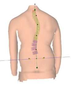
Because of this automatic measuring it is more accurate than the old way of calculating and measuring by hand. The numbers we get are accurate and clinically useful and help us to compare to previous scans.
One of the crucial areas measured is the pelvis. Information on position of the pelvis can then help us determine the possibility of one leg being longer than another, or it being out of alignment. These measurements happen in seconds without user error.
Three Dimensional Measurements
One big advantage with topography is we can measure all 3 planes and create 3D measurements and imaging. This allows for measurements of rotation which are crucial in the assessment of scoliosis.
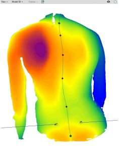
3D measurements also allow us to see all planes with only taking one image. With an x-ray, you need images from the front or back and then from the side to see 2 planes. To get full 3 dimensional imaging, you need to have additional imaging or special software to interpret the different views. Therefore 3D imaging using x-ray is generally not available.
Dynamic Correction Imaging
One of the coolest things with topography is to image the back while performing scoliosis, or posture, corrections. Since we don’t have to worry about radiation since we are just using light, we can do this as much as we want.
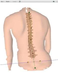
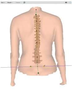
For my patients, this is invaluable. With these images while they are trying to perform their correction exercises, we can know if they are doing them correctly. If we see something off, it is easy to see and correct.
Now, there are some limitation of Surface Topography as well.
Limitations
It Doesn’t Actually “See” the Spine
Because we are only seeing the surface of the skin with lights projected on it, we are not actually seeing the spine. This allows for great measurements, like mentioned above, but does not allow us to see the actual spine.
This is why we cannot measure of Cobb angle in Scoliosis. To measure this angle, you need to see where each vertebrae is, not just a graphic representation.
With not seeing the spine, we also cannot make judgements on specific spine deformity such as congenital malformations of the spine, or fractures. This is why we usually have an x-ray to consult prior to treatment.
Not Completely Agreeing with X-ray Measurements
Due to the above limitation of it not seeing the spine, when the DIERS Formetric estimates the scoliosis angle, there is a mild error rate. In the research, this error can be up to 7 degrees, with lumbar curves being underestimated the most.
Postural measurements of kyphosis and lordosis are very comparable to x-ray however, and are very useful.
It may seem that not agreeing with x-ray is a huge limitation for Surface Topography, but that is not true. The purpose of this kind of measurement is to get more information about the back in 3D. Along with that, one of the main reasons for scanning with topography is to identify changes in the spine. This means we want to compare one scan to another scan down the road…not to an x-ray.
The accuracy of comparing these scans to each other is very high, even if the agreement with an x-ray is low. This makes it a very good tool for detecting progression.
It Can Be Uncomfortable
To get an unobstructed view of the back, the subject being scanned cannot have anything on the back. This usually mean shirt, and the pant line needs to be down lower than the belt line.
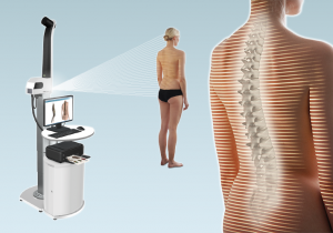
We address this by having a staff member of the same gender take the picture, and having the patient wear a gown or keep their shirt on until the machine is ready. Then a quick relaxed picture is taken. The person if faced away from the camera at all times to maintain privacy.
Who Could Benefit?
There are many conditions and diagnoses I use this type of imaging with. The following are common problems and diagnoses we scan.
- Scoliosis
- Scheurmanns Kyphosis
- Postural Kyphosis
- Sway Back
- Sacroiliac (SI) Joint Dysfunction
- Osteoporosis (to monitor posture)
- Leg Length Discrepancies
- Flat Back
- Poor Posture
- Headaches
- Shoulder Pain (to identify scapular issues)
- Spondylolisthesis
- Chronic Back Pain
If you have any of the issues listed above, and would like to learn if Surface Topography is right for you, please contact me by either calling (801)980-0860, or email me at david@aligntherapyutah.com.
At Align Therapy we are committed to providing the BEST and most Effective treatment for spinal deformities and back pain. We have invested in the most advanced tools and techniques to help our patients achieve their goals.


